
Enabling technology
Surgical Microscope
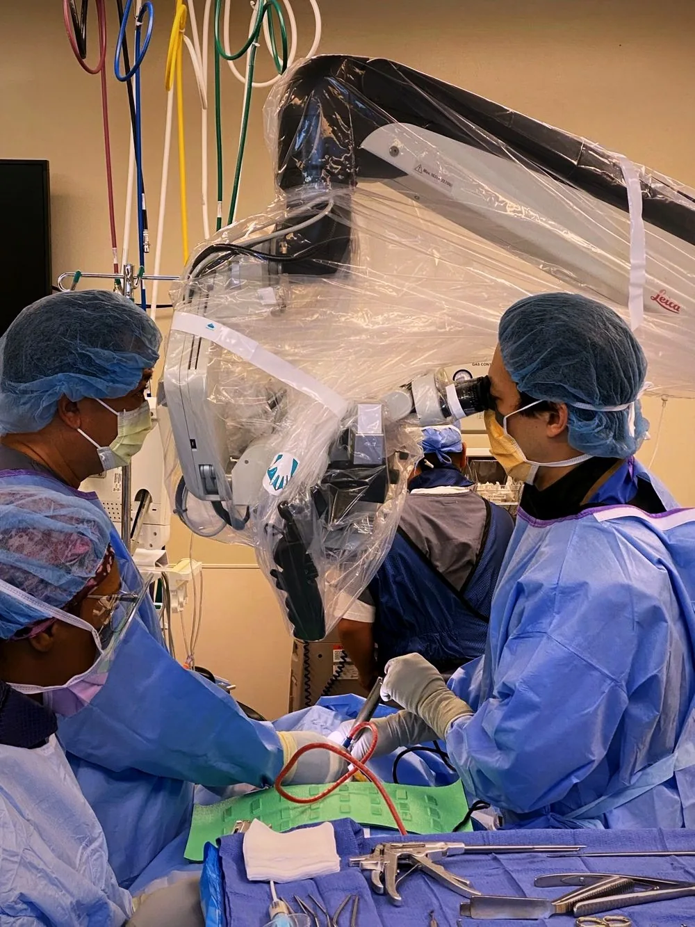
The operative microscope magnifies the surgical field many times over and focuses light exactly where it is needed, letting the surgeon distinguish nerve, disc, and bone in fine detail.
What it is
The surgical microscope is an optical instrument that brings high magnification and a brightly illuminated, enlarged view of the surgical field directly to the surgeon's eyes. It is a fixture of modern spinal decompression — from lumbar microdiscectomy to cervical foraminotomy.
Under the microscope, the distinction between compressed nerve tissue and the disc or bone pressing on it becomes far clearer than with the unaided eye, allowing the surgeon to work millimeter by millimeter.
How it supports precision
During decompression, the goal is to remove exactly the tissue causing compression while preserving everything else — nerve, facet joint, and stabilizing structures. Magnification makes that selective removal possible, and focused illumination eliminates the shadows that can hide delicate anatomy.
For patients, the practical payoff of this precision is a thoroughly decompressed nerve through a small, carefully managed corridor — consistent with the minimally invasive philosophy of doing the complete job with less disruption.
Its limits
The microscope shows what is in front of it — it cannot see around corners, through bone, or beyond the exposure that has been created. Its narrow field of view means the surgeon must actively survey the anatomy rather than assume what is nearby.
Like any tool, it amplifies both skill and judgment: it reveals detail, but the plan for what to remove and what to preserve is made by the surgeon, not the optics.
Initial consultation or second opinion?
Bring your imaging and your questions. Dr. Choi reviews the problem carefully and explains the options — including whether surgery is even the right next step.